Pneumothorax Chest X Ray / Chest X-ray showing large right pneumothorax with ... - In this video, you'll learn how to identify when radiological pleura is abnormal and the key signs to look out for when trying to diagnose a pneumothorax.
Pneumothorax Chest X Ray / Chest X-ray showing large right pneumothorax with ... - In this video, you'll learn how to identify when radiological pleura is abnormal and the key signs to look out for when trying to diagnose a pneumothorax.. A spontaneous pneumothorax, also referred to as a primary pneumothorax, occurs in the absence of a traumatic injury to the chest or a known lung disease. Reduction in lung markings in the apices (erect). But clinical correlation is needed for the final diagnosis (poor hemodynamic situation). Rib detail images may be taken to delineate bone. A secondary (also termed complicated) pneumothorax occurs due to an underlying condition.
You ascertain that this film is that of your patient's. The pneumothorax is an abnormal accumulating of. In those with secondary spontaneous pneumothorax due to these images are a random sampling from a bing search on the term chest xray in pneumothorax. click on the image (or right click) to open the. A collection of air within the pleural space between the lung (visceral pleura) and the chest wall (parietal pleura) that can lead to partial or. If you'd like to support us and get something great in return, check out our osce checklist if a tension pneumothorax is suspected clinically (shortness of breath and tracheal deviation) then immediate intervention should be performed without waiting for imaging as.
Pneumothorax | Chest X-Ray - MedSchool from medschool.co Top tips for pneumothorax aspiration (thoracocentesis). Check the full list of possible causes and conditions now! Transparent films, which can be stimulated by laser beams after x‐ray irradiation, have been searched to improve the spatial resolution of digital x‐ray imaging sensors. Quantification of pneumothorax size on chest radiographs using interpleural distances: Pneumothorax occurs when air leaks from inside of the lung to the space between the lung and the chest wall. Pneumothorax due to apical blebs. A pneumothorax is an abnormal collection of air in the pleural space between the lung and the chest wall. Diagnosis of pneumothorax by radiography and ultrasonography:
In those with secondary spontaneous pneumothorax due to these images are a random sampling from a bing search on the term chest xray in pneumothorax. click on the image (or right click) to open the.
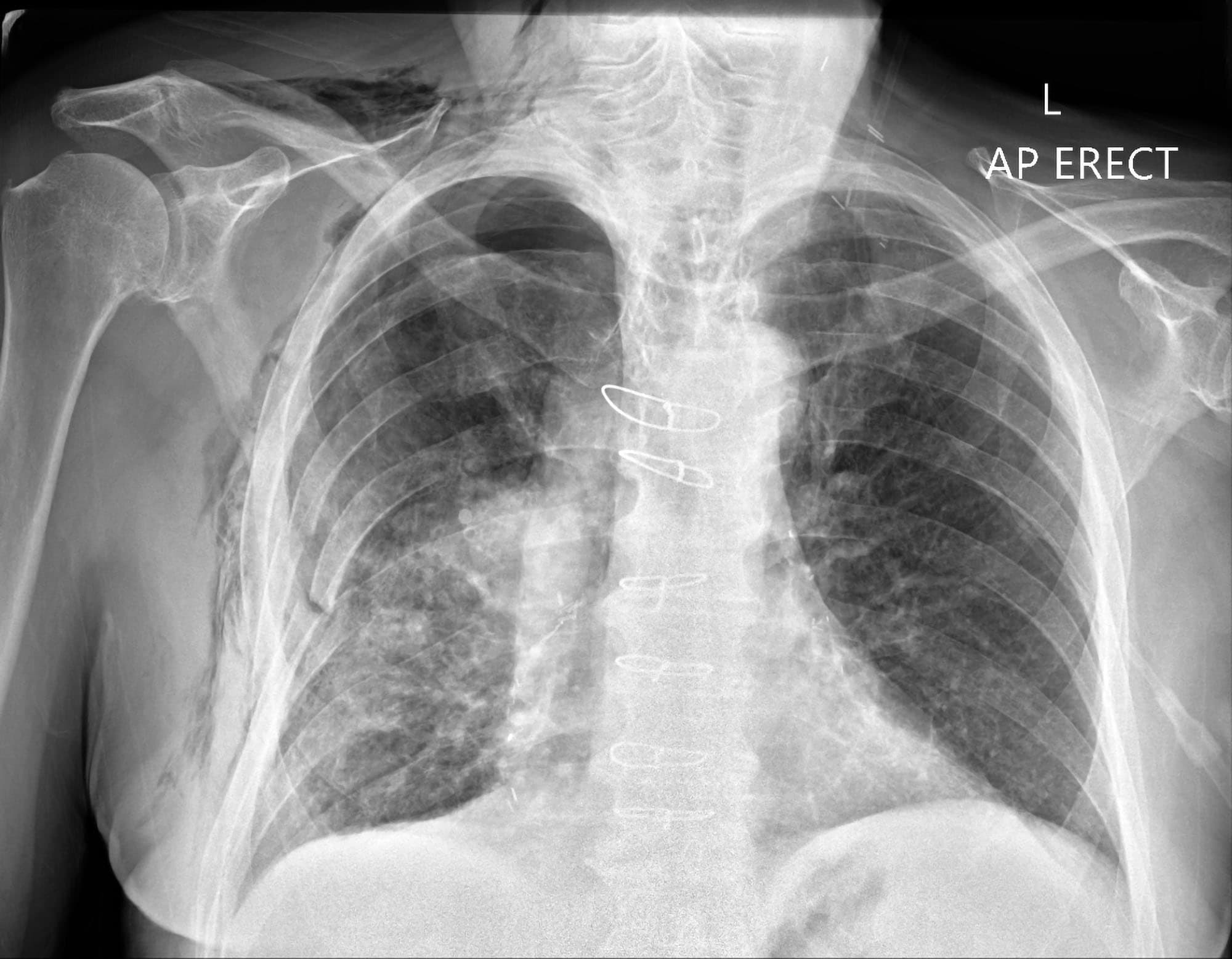
In fact every radiologst should be an expert in chest film reading. We see a large pneumothorax, which is causing collapse of the left lung and displacement of the mediastinal structures to the right. A diagnosis of tension pneumothorax is primarily a clinical one because the condition itself is an emergency and warrants initiation of an. If pneumothorax remains greater than 2cm, proceed to a further attempt at aspiration. Pneumothorax can occur spontaneously or result from trauma or medical procedures. A spontaneous pneumothorax, also referred to as a primary pneumothorax, occurs in the absence of a traumatic injury to the chest or a known lung disease. Quantification of pneumothorax size on chest radiographs using interpleural distances: Indicated where chest xray cannot distinguish bleb in copd from pneumothorax. Top tips for pneumothorax aspiration (thoracocentesis). Regression analysis based on volume measurements from helical ct. A pneumothorax is an abnormal collection of air in the pleural space between the lung and the chest wall. A chest ct scan can provide information about the underlying cause such as a bullae in cases of spontaneous pneumothorax. But clinical correlation is needed for the final diagnosis (poor hemodynamic situation).
In this video, you'll learn how to identify when radiological pleura is abnormal and the key signs to look out for when trying to diagnose a pneumothorax. Rib detail images may be taken to delineate bone. We see a large pneumothorax, which is causing collapse of the left lung and displacement of the mediastinal structures to the right. A pneumothorax refers to the presence of gas or air in the pleural space. The pneumothorax is an abnormal accumulating of.
Tension pneumothorax and the "forbidden CXR" -- McRoberts ... from emj.bmj.com If discharging a patient with a residual pneumothorax ensure they are aware of this and that they know. In those with secondary spontaneous pneumothorax due to these images are a random sampling from a bing search on the term chest xray in pneumothorax. click on the image (or right click) to open the. A collection of air within the pleural space between the lung (visceral pleura) and the chest wall (parietal pleura) that can lead to partial or. A spontaneous pneumothorax, also referred to as a primary pneumothorax, occurs in the absence of a traumatic injury to the chest or a known lung disease. Rib detail images may be taken to delineate bone. Pneumothorax can occur spontaneously or result from trauma or medical procedures. Small pneumothoraces may resorb spontaneously, but larger defects usually pneumothorax: Diagnosis of pneumothorax by radiography and ultrasonography:
Top tips for pneumothorax aspiration (thoracocentesis).
Transparent films, which can be stimulated by laser beams after x‐ray irradiation, have been searched to improve the spatial resolution of digital x‐ray imaging sensors. But clinical correlation is needed for the final diagnosis (poor hemodynamic situation). You ascertain that this film is that of your patient's. If you'd like to support us and get something great in return, check out our osce checklist if a tension pneumothorax is suspected clinically (shortness of breath and tracheal deviation) then immediate intervention should be performed without waiting for imaging as. In this video, you'll learn how to identify when radiological pleura is abnormal and the key signs to look out for when trying to diagnose a pneumothorax. Pneumothorax can occur spontaneously or result from trauma or medical procedures. Traumatic pneumothorax detection with thoracic us: Pneumothorax occurs when air leaks from inside of the lung to the space between the lung and the chest wall. Pneumothorax due to apical blebs. Indicated where chest xray cannot distinguish bleb in copd from pneumothorax. Quantification of pneumothorax size on chest radiographs using interpleural distances: In fact every radiologst should be an expert in chest film reading. We see a large pneumothorax, which is causing collapse of the left lung and displacement of the mediastinal structures to the right.
If pneumothorax remains greater than 2cm, proceed to a further attempt at aspiration. The pneumothorax is an abnormal accumulating of. In this video, you'll learn how to identify when radiological pleura is abnormal and the key signs to look out for when trying to diagnose a pneumothorax. The lungs can be seen to reach the inner edge of the thoracic wall (arrows). Indicated where chest xray cannot distinguish bleb in copd from pneumothorax.
Pneumothorax Chest X-Ray | LE PNEUMOTHORAX | X-Ray | Pinterest from s-media-cache-ak0.pinimg.com If you'd like to support us and get something great in return, check out our osce checklist if a tension pneumothorax is suspected clinically (shortness of breath and tracheal deviation) then immediate intervention should be performed without waiting for imaging as. We see a large pneumothorax, which is causing collapse of the left lung and displacement of the mediastinal structures to the right. Rib detail images may be taken to delineate bone. In this video, you'll learn how to identify when radiological pleura is abnormal and the key signs to look out for when trying to diagnose a pneumothorax. Top tips for pneumothorax aspiration (thoracocentesis). Pneumothorax occurs when air leaks from inside of the lung to the space between the lung and the chest wall. A collection of air within the pleural space between the lung (visceral pleura) and the chest wall (parietal pleura) that can lead to partial or. Small pneumothoraces may resorb spontaneously, but larger defects usually pneumothorax:
But clinical correlation is needed for the final diagnosis (poor hemodynamic situation).
Of the 54 chest roentgenograms, 52 were obtained in asymptomatic patients, with no suspicion of pneumothorax. Quantification of pneumothorax size on chest radiographs using interpleural distances: Small pneumothoraces may resorb spontaneously, but larger defects usually pneumothorax: It is considered a simple pneumothorax when there isn't any mediastinal shift to. Regression analysis based on volume measurements from helical ct. Top tips for pneumothorax aspiration (thoracocentesis). A chest ct scan can provide information about the underlying cause such as a bullae in cases of spontaneous pneumothorax. A secondary (also termed complicated) pneumothorax occurs due to an underlying condition. Traumatic pneumothorax detection with thoracic us: If you'd like to support us and get something great in return, check out our osce checklist if a tension pneumothorax is suspected clinically (shortness of breath and tracheal deviation) then immediate intervention should be performed without waiting for imaging as. Transparent films, which can be stimulated by laser beams after x‐ray irradiation, have been searched to improve the spatial resolution of digital x‐ray imaging sensors. You ascertain that this film is that of your patient's. Also, the presence of air outside normal lung airways and movement or shifting.
You have just read the article entitled Pneumothorax Chest X Ray / Chest X-ray showing large right pneumothorax with ... - In this video, you'll learn how to identify when radiological pleura is abnormal and the key signs to look out for when trying to diagnose a pneumothorax.. You can also bookmark this page with the URL : https://nikolaussan.blogspot.com/2021/05/pneumothorax-chest-x-ray-chest-x-ray.html
Share Awesome
Belum ada Komentar untuk "Pneumothorax Chest X Ray / Chest X-ray showing large right pneumothorax with ... - In this video, you'll learn how to identify when radiological pleura is abnormal and the key signs to look out for when trying to diagnose a pneumothorax."


Belum ada Komentar untuk "Pneumothorax Chest X Ray / Chest X-ray showing large right pneumothorax with ... - In this video, you'll learn how to identify when radiological pleura is abnormal and the key signs to look out for when trying to diagnose a pneumothorax."
Posting Komentar