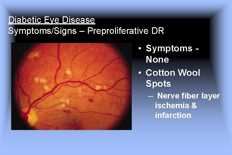
Cotton Wool Spots Symptoms - Several Cotton Wool Spots Typical Of Hiv Retinopathy Are Seen In This Download Scientific Diagram : White spots on retinal surface caused by microinfarction.
Cotton Wool Spots Symptoms - Several Cotton Wool Spots Typical Of Hiv Retinopathy Are Seen In This Download Scientific Diagram : White spots on retinal surface caused by microinfarction.. Hypertension, connective tissue disease, or aids) causing hypoxia (oxygen deficiency) in the nerve fibre layer … the new mediacal dictionary. Various degree of blurry vision. Cotton wool spots symptom checker: Causes are hypertension, diabetes, hiv, lupus, severe anemia or thrombocytopenia, hypercoagulable states, connective tissue disorders, viruses. How is cotton wool spots diagnosed & treated.
Possible causes of cotton wool spots (medical symptom) cotton wool spots are an abnormal finding on fundoscopic exam of the retina of the eye this video. Hypertensive retinopathy is a hallmark of high blood pressure. They appear as fluffy white patches on the retina. The cotton wool appearance is a plain film sign of paget disease and results from thickened, disorganized trabeculae which lead to areas of sclerosis in a previously lucent area of bone, typically the skull. When volume reaches 80ml will see acute signs and symptoms**.
Can You Spot The Problem from www.reviewofoptometry.com Symptoms cotton wool spots are asymptomatic. They are caused by damage to nerve fibers and are a result of accumulations of axoplasmic material within the nerve fiber layer. This section shows a full list of all the diseases and conditions listed as a possible cause of cotton wool spots in our database from various sources. Cotton wool spots have a differential diagnosis list about a mile long. Cotton wool spots are an abnormal finding on funduscopic exam of the retina of the eye. Primary workup sphygmomanometry (high blood pressure) fbs/hba1c (diabetes) ana (sle. In otherwise healthy patients, the observance of a cotton wool spot (cws) is not considered normal. Why cotton wool spots should not be regarded as retinal nerve fibre layer infarcts.
Signs cotton wool spots appear on fundoscopy as white, fluffy lesions with hazy or feathered edges in the superficial retina.
Diabetes and hypertension are the two most common diseases that cause these spots, and the best treatment would be to treat the underlying disease. Amongst the causes are lupus and anemia and untreated hypertension. Verwenden sie den chatbot, um ihre suche weiter zu verfeinern. Cotton wool spots are an abnormal finding on funduscopic exam of the retina of the eye. The cotton wool appearance is a plain film sign of paget disease and results from thickened, disorganized trabeculae which lead to areas of sclerosis in a previously lucent area of bone, typically the skull. They appear as fluffy white patches on the retina. Mögliche ursachen sind unter anderem präeklampsie. A single cotton wool spot in one eye can be the earliest ophthalmoscopic finding in diabetic or hypertensive retinopathy. Cotton wool spot right eye found on annual eye exam. Possible causes of cotton wool spots (medical symptom) cotton wool spots are an abnormal finding on fundoscopic exam of the retina of the eye this video. They are caused by damage to nerve fibers and are a result of accumulations of axoplasmic material within the nerve fiber layer. They appear as fluffy white patches on the retina. Symptoms of hypertensive retinopathy form arteriolar narrowing, while the other.
Cotton wool spots are an abnormal finding on funduscopic exam of the retina of the eye. They appear as fluffy white patches on the retina. What are causes & symptoms of cotton wool spots. Cotton wool spots can be defined as the abnormal findings that are identified on the regular examination of the retina (fundoscopic examination). When volume reaches 80ml will see acute signs and symptoms**.
Hypertensive Retinopathy Symptoms Causes And Treatments from i0.wp.com Mögliche ursachen sind unter anderem präeklampsie. Formation of cotton wool spots (cws) is one of the important clinical signs that are seen at advanced stages in hr. Hypertension, diabetes, ocular ischemic syndrome (cotton wool spots is less common), retinal vein occlusion, anemia. They appear as fluffy white patches on the retina. The cotton wool appearance is a plain film sign of paget disease and results from thickened, disorganized trabeculae which lead to areas of sclerosis in a previously lucent area of bone, typically the skull. Diabetes and hypertension are the two most common diseases that cause these spots, and the best treatment would be to treat the underlying disease. Detailed analysis of 8 causes of cotton wool spots symptom, alternative diagnoses and related symptoms. They are caused by damage to nerve fibers and are a result of accumulations of axoplasmic material within the nerve fiber layer.
They are most commonly seen in patients with diabetes mellitus or systemic hypertension.
Cotton wool spots are an abnormal finding on funduscopic exam of the retina of the eye. Mögliche ursachen sind unter anderem präeklampsie. When volume reaches 80ml will see acute signs and symptoms**. They are caused by damage to nerve fibers and are a result of accumulations of axoplasmic material within the nerve fiber layer. Usually do not produce vision loss unless large or near fovea. Also, if the patient suffers from an underlying condition, such as diabetes or hypertension, he/she will present the characteristic symptoms for the. Amongst the causes are lupus and anemia and untreated hypertension. Cotton wool spots autonomic nervous systems physical exam findings rapid acting insulin fasting blood glucose. They are caused by damage to nerve fibers and are a result of accumulations of axoplasmic material within the nerve fiber layer. Signs cotton wool spots appear on fundoscopy as white, fluffy lesions with hazy or feathered edges in the superficial retina. In otherwise healthy patients, the observance of a cotton wool spot (cws) is not considered normal. Cotton wool spot right eye found on annual eye exam. Symptoms of hypertensive retinopathy form arteriolar narrowing, while the other.
Detailed analysis of 8 causes of cotton wool spots symptom, alternative diagnoses and related symptoms. Also, if the patient suffers from an underlying condition, such as diabetes or hypertension, he/she will present the characteristic symptoms for the. They are caused by damage to nerve fibers and are a result of accumulations of axoplasmic material within the nerve fiber layer. Many articles in the literature have reported that they are seen in retinopathies due to a whole host of conditions. They are caused by damage to nerve fibers and are a result of accumulations of axoplasmic material within the nerve fiber layer.
Diabetic Eye Disease Evan Jake Waxman Md Ph from slidetodoc.com They appear as fluffy white patches on the retina. Hypertension, diabetes, ocular ischemic syndrome (cotton wool spots is less common), retinal vein occlusion, anemia. Cotton wool spots causes can be categorized into: They are caused by damage to nerve fibers and are a result of accumulations of axoplasmic material within the nerve fiber layer. Various degree of blurry vision. Formation of cotton wool spots (cws) is one of the important clinical signs that are seen at advanced stages in hr. These usually point towards some systemic problem. These sclerotic patches are poorly def.
They are caused by damage to nerve fibers and are a result of accumulations of axoplasmic material within the nerve fiber layer.
Symptoms cotton wool spots are asymptomatic. Verwenden sie den chatbot, um ihre suche weiter zu verfeinern. Hypertension, connective tissue disease, or aids) causing hypoxia (oxygen deficiency) in the nerve fibre layer … the new mediacal dictionary. Also, if the patient suffers from an underlying condition, such as diabetes or hypertension, he/she will present the characteristic symptoms for the. These usually point towards some systemic problem. They appear as fluffy white patches on the retina. Hamamoto and ronakorn panjaphongse}, journal={2018 11th biomedical. Various degree of blurry vision. Hypertensive retinopathy is a hallmark of high blood pressure. Cotton wool spots autonomic nervous systems physical exam findings rapid acting insulin fasting blood glucose. Hypertension, diabetes, ocular ischemic syndrome (cotton wool spots is less common), retinal vein occlusion, anemia. Many articles in the literature have reported that they are seen in retinopathies due to a whole host of conditions. Diabetes and hypertension are the two most common diseases that cause these spots, and the best treatment would be to treat the underlying disease.
You have just read the article entitled Cotton Wool Spots Symptoms - Several Cotton Wool Spots Typical Of Hiv Retinopathy Are Seen In This Download Scientific Diagram : White spots on retinal surface caused by microinfarction.. You can also bookmark this page with the URL : https://nikolaussan.blogspot.com/2021/06/cotton-wool-spots-symptoms-several.html
Share Awesome
Belum ada Komentar untuk "Cotton Wool Spots Symptoms - Several Cotton Wool Spots Typical Of Hiv Retinopathy Are Seen In This Download Scientific Diagram : White spots on retinal surface caused by microinfarction."

Belum ada Komentar untuk "Cotton Wool Spots Symptoms - Several Cotton Wool Spots Typical Of Hiv Retinopathy Are Seen In This Download Scientific Diagram : White spots on retinal surface caused by microinfarction."
Posting Komentar